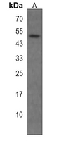
Western blot analysis of STAMBPL1 expression in Hela (A) whole cell lysates.
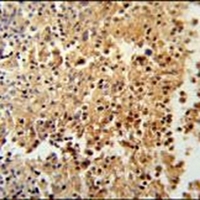
Immunohistochemical analysis of STAMBPL1 staining in human spleen formalin fixed paraffin embedded tissue section.
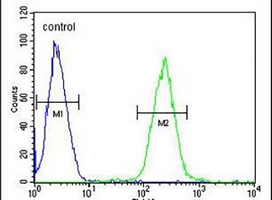
Flow cytometric analysis of CEM cells using Anti-STAMBPL1 Antibody. The cells were fixed with 2% paraformaldehyde (10 min) and then permeabilized with 90% methanol for 10 min. The cells were incubated in 2% bovine serum albumin to block non-specific protein-protein interactions followed by the antibody at 37 °C for 60 min. The secondary antibody Goat Anti-Rabbit IgG (H&L) - AF488 was incubated at 37 °C for 40 min. Isotype control antibody (blue line) was used under the same condition.