中国 EN 400-660-9091

联系我们

电话:18013895086

电话:025-68037686

地址:江苏生命科技创新园F6幢1层

订购:nanjing03@biogot.com

服务:biorase01@biogot.com

合作:lvyun@biogot.com

支持:may@biogot.com

LUC7L polyclonal antibody

货号: BS32436
价格 1140
货期 现货(1-2个工作日)
25ul 50ul 100ul 200ul
No. :BS32436
Product Name :LUC7L polyclonal antibody
Swiss-Prot :Q9NQ29
Host :Rabbit
Reactivity :Human
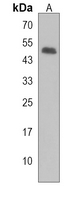
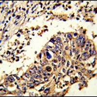
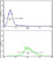
Applications :WB, IHC, FC
Application_all :WB (1/500 - 1/1000), IHC (1/50 - 1/200), FC (1/10 - 1/50)
Background :Putative RNA-binding protein Luc7-like 1: May bind to RNA via its Arg/Ser-rich domain
Product :1mg/ml in 0.42% Potassium phosphate, 0.87% Sodium chloride, pH 7.3, 30% glycerol, and 0.01% sodium azide.
Purification&Purity :The antibody was purified by immunogen affinity chromatography.
Storage&Stability :Store at 4°C short term. Aliquot and store at -20°C long term. Avoid freeze-thaw cycles.
Specificity :Recognizes endogenous levels of LUC7L protein.
BiowMW :~43.7 kDa
Note :For research use only, not for use in diagnostic procedure.
Alternative Name :LUC7L1; Putative RNA-binding protein Luc7-like 1; Putative SR protein LUC7B1; SR+89
Immunogen :A synthetic peptide encompassing a sequence within the C-terminal region of human LUC7L.
Conjugate :Unconjugated
Modification :Unmodification
Western blot analysis of CD222 expression in Jurkat (A) whole cell lysates.
Immunohistochemical analysis of CD222 staining in human colon formalin fixed paraffin embedded tissue section. The section was pre-treated using heat mediated antigen retrieval with sodium citrate buffer (pH 6.0). The section was then incubated with the antibody at room temperature and detected using an HRP conjugated compact polymer system. DAB was used as the chromogen. The section was then counterstained with haematoxylin and mounted with DPX.
Immunofluorescent analysis of CD222 staining in Jurkat cells. Formalin-fixed cells were permeabilized with 0.1% Triton X-100 in TBS for 5-10 minutes and blocked with 3% BSA-PBS for 30 minutes at room temperature. Cells were probed with the primary antibody in 3% BSA-PBS and incubated overnight at 4 °C in a hidified chamber. Cells were washed with PBST and incubated with an AF488-conjugated secondary antibody (green) in PBS at room temperature in the dark. Phalloidin - AF594 was used to stain Actin filaments (red). DAPI was used to stain the cell nuclei (blue).


关注我们

400-660-9091

电话:18013895086

地址:江苏生命科技创新园F6幢1层

订购:nanjing03@biogot.com

服务:biorase01@biogot.com

合作:lvyun@biogot.com

支持:may@biogot.com