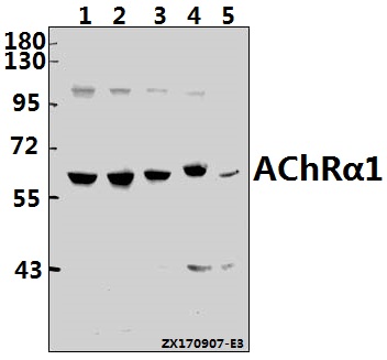
Western blot (WB) analysis of AChRα1 (E217) pAb at 1:500 dilution
Lane1:HepG2 whole cell lysate(40ug)
Lane2:A549 whole cell lysate(40ug)
Lane3:PC3 whole cell lysate(40ug)
Lane4:CT26 whole cell lysate(40ug)
Lane5:H9C2 whole cell lysate(40ug)
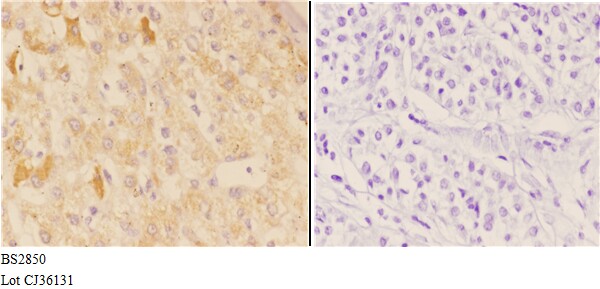
Immunohistochemistry (IHC) analyzes of AChRα1 (E217) pAb bin paraffin-embedded human liver carcinoma tissue at 1:50,showing membrnae and cytoplasmic staining.Negative control (the right)Using PBS instead of primary antibody, secondary antibody is Goat Anti-Rabbit IgG-biotin followed by avidin-peroxidase.
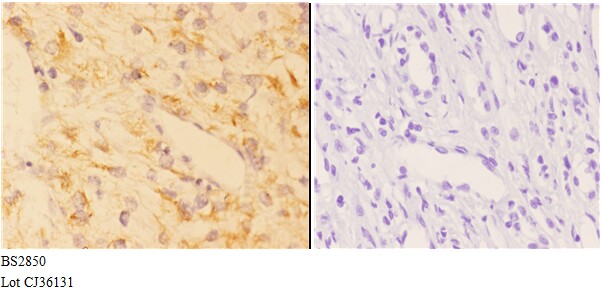
Immunohistochemistry (IHC) analyzes of AChRα1 (E217) pAb bin paraffin-embedded human kidney carcinoma tissue at 1:50,showing membrnae and cytoplasmic staining.Negative control (the right)Using PBS instead of primary antibody, secondary antibody is Goat Anti-Rabbit IgG-biotin followed by avidin-peroxidase.