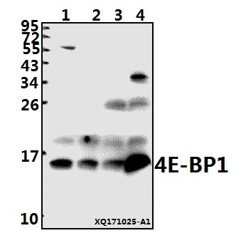
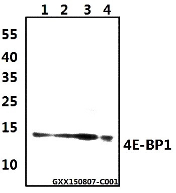
Western blot (WB) analysis of 4E-BP1 (G39) pAb at 1:500 dilution
Lane1:HEK293T whole cell lysate(40ug)
Lane2:PC3 whole cell lysate(40ug)
Lane3:The Ovary tissue lysate of Rat(40ug)
Lane4:The Embryo tissue lysate of Mouse(40ug)
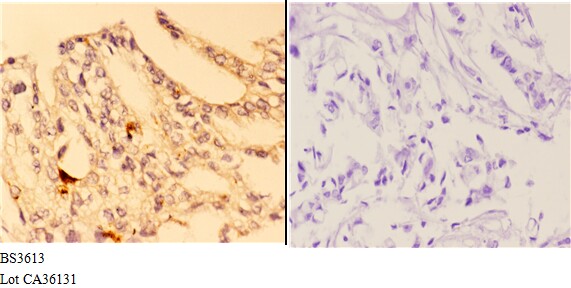
Immunohistochemistry (IHC) analyzes of 4E-BP1 (G39) pAb in paraffin-embedded human breast carcinoma tissue at 1:50,showing cytoplasmic staining.Negative control (the right)Using PBS instead of primary antibody, secondary antibody is Goat Anti-Rabbit IgG-biotin followed by avidin-peroxidase.
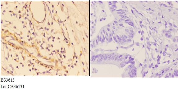
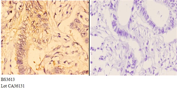
Immunohistochemistry (IHC) analyzes of 4E-BP1 (G39) pAb in paraffin-embedded human colon carcinoma tissue at 1:50,showing cytoplasmic staining.Negative control (the right)Using PBS instead of primary antibody, secondary antibody is Goat Anti-Rabbit IgG-biotin followed by avidin-peroxidase.
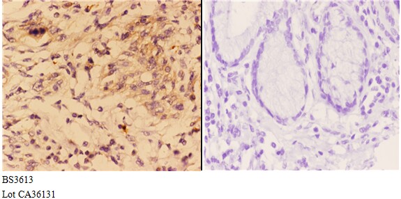
Immunohistochemistry (IHC) analyzes of 4E-BP1 (G39) pAb in paraffin-embedded human esophageal carcinoma tissue at 1:50,showing cytoplasmic staining.Negative control (the right)Using PBS instead of primary antibody, secondary antibody is Goat Anti-Rabbit IgG-biotin followed by avidin-peroxidase.